Cruciate injuries and repairs
Cruciate rupture or injury is by far the most common hind leg lameness issue in dogs. It is also our most common orthopaedic repair. The link below is a good read and explains the condition
https://www.fitzpatrickreferrals.co.uk/orthopaedic/cranial-cruciate-ligament-injury/
Please note that Flynn Vets is not affiliated with Fitzpatrick Referrals.
At Flynnvets in Belfast and Ballynahinch our Orthopaedic Surgeon Dan Flynn has repaired cruciate ruptures in a number of different ways over the years. We are constantly trying to improve surgery techniques to get the best recovery and long term outcome for our patients.
What are the main types of dog cruciate ligament surgery?
The main types of surgery used for repair of cruciate ligaments are:
Lateral Suture technique – no longer used at Flynnvets
Modified Maquet procedure (MMP) – no longer used at Flynnvets
Tibial Tuberosity Advancement (TTA) – Very occasionally used
Cranial Closing Wedge Osteotomy (CCWO) – (usually mainly in dogs of 3.5kg-25kg)
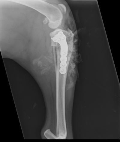
Tibial Plateau Leveling Osteotomy (TPLO) – (used for dogs of 10kg-80kg)
What type of dog cruciate ligament surgery do we use?
We have progressed through all of these techniques over the past decade and now use the TPLO as our first option, except for very small dogs where the CCWO remains our procedure of choice.
The video (right) is Pippa who is 3.5 kgs and 9 years old. It shows her 7 days after we did a CCWO on her right hind leg. Her recovery is amazing, especially considering this is her second operation – she had her left leg repaired just 3 months earlier. When we see how well they do post surgery we have no hesitation in recommending the procedure, even in older dogs.
We use Securos Surgical PAX Polyaxial Locking Plates System for our implants: this is new technology in orthopaedics and provides more robust security in screw placement, leading to reduced complications and improved outcomes.
Pippa - day 7 after surgery
Case Study
Chucky, a 57kg Rottweiler also underwent a TPLO procedure with us recently. The X-ray (right) was taken during his surgery, which involved an 8mm rotation to the tibia. You can clearly see where a section has been cut from the top of the tibia and displaced to alter the angle of the joint.

